Health is wealth. For every one of us health is more important than any other thing. Good food, ans meditation too will keep us healthy.
Today with the evolution of technology medical field has also improved and have achieved important milestone using the technology. Human body is made of some sensitive tissues, if anything happens to that tissues or bones it is not easy to identify. However, using one technology we can easily identify problems in our inner tissues or bones. That is X-ray.


X-ray radiation, or, much less commonly, X-radiation, is a penetrating form of high-energy electromagnetic radiation. An X-ray is produced when a negatively charged electrode is heated by electricity and electrons are released, thereby producing energy. That energy is directed toward a metal plate, or anode, at high velocity and an X-ray is produced when the energy collides with the atoms in the metal plate.
X-rays are used in ways such as checking for broken bones, detecting certain kinds of diseases, identification of some metals, and ascertaining the locations of weak points in steel.
The X-ray was accidentally discovered by Wilhelm Roentgen, a professor of physics in Wurzburg, Bavaria in 1895 during an experiment. In 1896, the first radiology department was established at the Glasgow Royal Infirmary. Soon, X-ray machines were being used for many things, such as diagnosing fractures in the bones and to locate bullets during WWI. This one discovery advanced the field of medicine in a very big way. To be able to see the structure inside a person without blood loss from an incision was revolutionary.
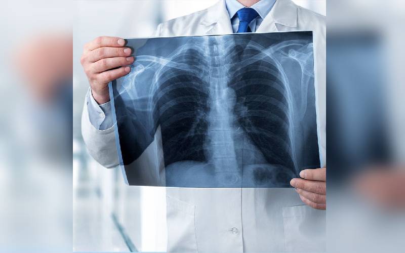
Medical x-rays are used to generate images of tissues and structures inside the body. If x-rays traveling through the body also pass through an x-ray detector on the other side of the patient, an image will be formed that represents the “shadows” formed by the objects inside of the body.
One type of x-ray detector is photographic film, but there are many other types of detectors that are used to produce digital images. The x-ray images that result from this process are called radiographs.
To create a radiograph, a patient is positioned so that the part of the body being imaged is located between an x-ray source and an x-ray detector. When the machine is turned on, x-rays travel through the body and are absorbed in different amounts by different tissues, depending on the radiological density of the tissues they pass through. Radiological density is determined by both the density and the atomic number (the number of protons in an atom’s nucleus) of the material being imaged.
Conversely, x-rays travel more easily through less radiologically dense tissues, such as fat, muscle, and air-filled cavities such as the lungs. These structures are displayed in shades of grey on a radiograph.
When used appropriately, the diagnostic benefits of x-ray scans significantly outweigh the risks. X-ray scans can diagnose possibly life-threatening conditions such as blocked blood vessels, bone cancer, and infections. However, x-rays produce ionising radiation—a form of radiation that has the potential to harm living tissue. This is a risk that increases with the number of exposures added up over the life of an individual. However, the risk of developing cancer from radiation exposure is generally small.
Current research of x-ray technology focuses on ways to reduce radiation dose, improve image resolution, and enhance contrast materials and methods.